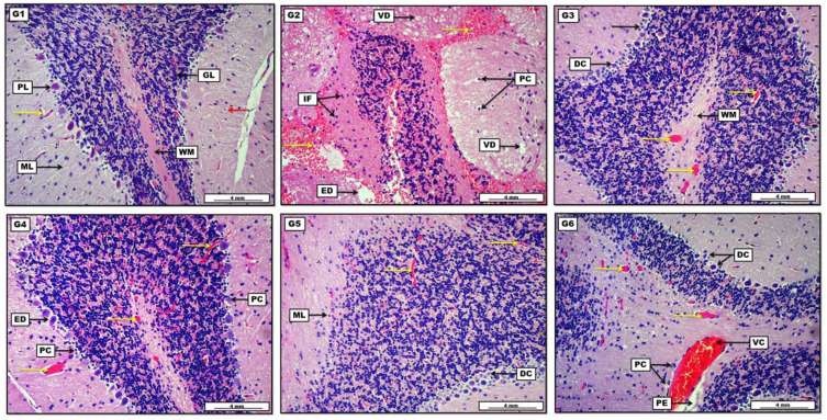
Figure 7.
Photomicrograph of brain tissue from groups; (G1): Display the intact white matter (WM), together with typically organized cerebral layers apparent with granular layer (GL), Purkinje cell layer (PL) and molecular layer (ML). The section also reveals slight capillary engorgement (yellow arrow) with few pyknotic cells (red arrow). (G2): Show the presence of diffuse pinkish edematous fluid (ED) mixed with a significant amount of inflammatory exudates (IF), together with diffuse areas of vascular hemorrhage (yellow arrows). The section also reveals many pyknotic cells (PC) as well as many (vacuolar degeneration). (G3): Display moderate vascular microthrombi (yellow arrows) distributed within the normal-appearing white matter (WM), together with the presence of some degenerative (DC) and pyknotic cells (black arrow) in the Purkinje cell layer. (G4): Show the presence of moderate capillary microthrombi (yellow arrows) in the areas of white matter and cerebral tissue. Moreover, the section reveals many pycnotic dark cells (PC) together with pericellular necrotic edema (ED). (G5): Display mildly distinct vascular microthrombi (yellow arrows), together with the incidence of some degenerative cells (DC), along with a few pyknotic debris within the cerebellar molecular layer (ML). (G6): Demonstrate many areas of severe vascular congestion (VC) and microthrombi capillary engorgement (yellow arrows), to be present with perivascular edema (PE) and perivascular coughing of inflammatory cells (PC). Additionally, the section shows the occurrence of many degenerative cells (DC) within the Purkinje cell layer. H&E. Scale bar: 4 mm.

