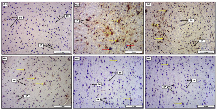
Figure 8.
Photomicrograph of brain tissue represents immunohistochemical staining with P53 antibody from groups; (G1): The section shows many weakly stained immunopositively nerve cells. (G2): Show the presence of large number of deeply brown stained immunopositively cells (G3): Demonstrate the presence of some moderately stained brownish immunopositively cells. (G4): Display the occurrence of many immunopositively neurons with moderate brownish chromatosis. (G5): Reveal little expression of weakly stained immunopositively nerve cells. (G6): Show the presence of tiny light-brown stained immunoreactive cells. Scale bar: 4 mm.

