Figure 7.

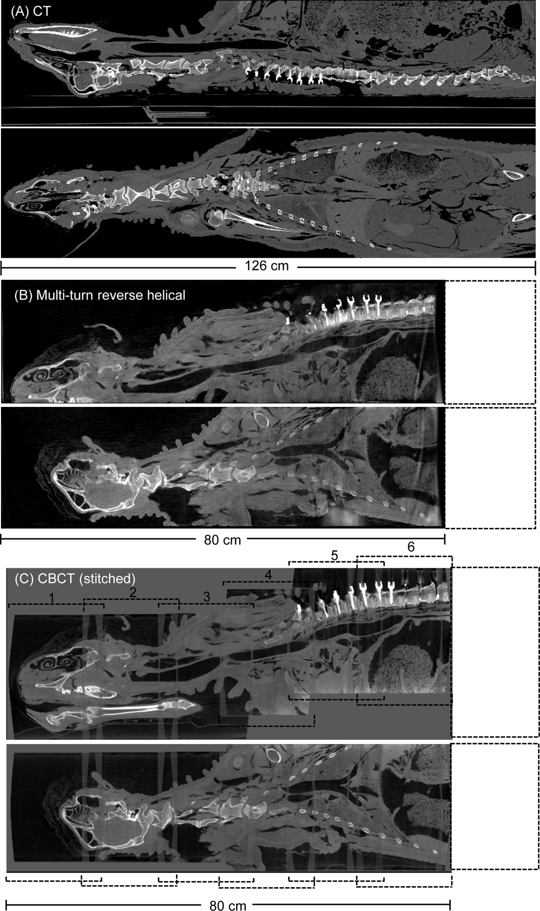
Post-operative reconstructed 3D images (sagittal and coronal view) of an ovine cadaver following a pedicle screw posterior fixation from T1-T12 acquired from (A) conventional CT, (B) the multi-turn reverse helical and (C), a series of conventional CBCT scans stitched together (numbered 1 through 6). All images reconstructed with metal artifact reduction (MAR) algorithms (in-built MAR for the conventional CT and CBCT). Intensity window display for the conventional CT scan [640, 800] HU. Intensity window display for the multi-turn reverse helical and stitched conventional CBCT scans [0.063, 0.025] mm−1.
