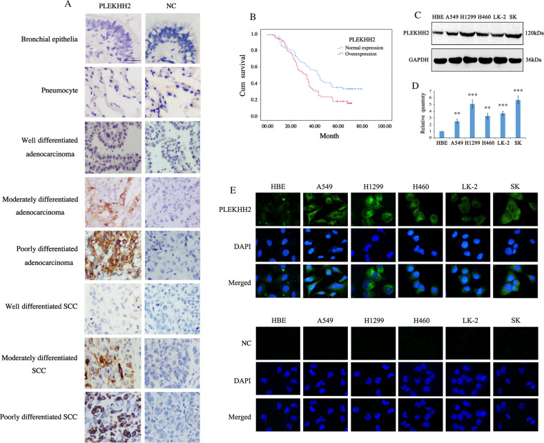
Fig. 1. PLEKHH2 is upregulated in non-small cell lung cancer.
A In normal bronchial and alveolar epithelia, negative PLEKHH2 staining was detected via immunohistochemical analysis. NSCLC samples showed cytoplasmic accumulation of PLEKHH2 protein, and it occurred more frequently in tumors with poorer differentiation (Rabbit IgG was used as a negative control; Scale bar, 50 μm). B Overexpression of PLEKHH2 correlates with poor patient survival. Among the 170 cases with follow-up information, Kaplan-Meier curves showed that patients with PLEKHH2 overexpression (94 cases) had a shorter survival time than those with normal PLEKHH2 expression (76 cases) (P = 0.014). C Western blot analysis of PLEKHH2 expression in lung cancer cells. Increased PLEKHH2 expression was detected in lung cancer cell lines compared to the immortalized bronchial epithelial cell line HBE. D PLEKHH2 protein levels were normalized to those of GAPDH. Statistical analysis showed increased PLEKHH2 expression in lung cancer cell lines compared to HBE cells. E Laser confocal microscopy images show that cytoplasmic PLEKHH2 expression was higher in lung cancer cells than in HBE cells. (PLEKHH2-CRISPR/CAS9 was used as negative control; Original magnification ×400). P < 0.05 indicates statistical significance, **P < 0.01, ***P < 0.001.

