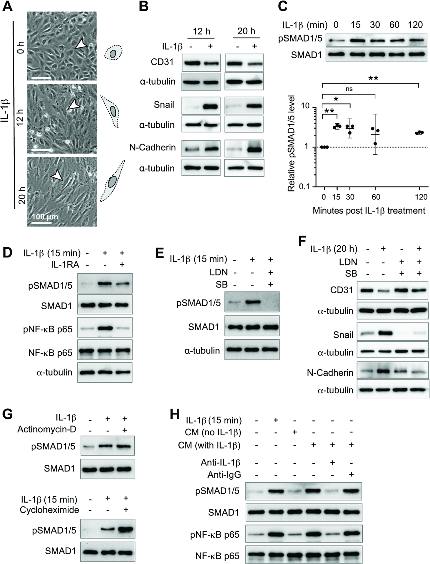
Figure 4. Crosstalk between IL-1β and ALK receptor signaling induces EndoMT in HBMECs.

(A) Morphological changes in HBMECs after IL-1β treatment. Quantification of changes in cell morphology is shown in Figure S7A.
For panels B-H, western blotting was used to assess protein levels.
(B) Endothelial cell marker CD31 and mesenchymal markers Snail and N-cadherin were assessed in HBMECs treated with IL-1β for 12 or 20 h.
(C) Levels of pSMAD1/5, an important ALK receptor downstream signaling molecule, were assessed in HBMECs at different time points following IL-1β treatment. One-way randomized block ANOVA with Dunnett’s correction for multiple comparisons following log10 transformation. *p<0.05, **p<0.01, ns: not significant. Geometric means and 95% confidence intervals are shown.
(D) HBMECs treated with or without IL-1RA were stimulated with IL-1β for 15 min. The level of pSMAD1/5 and pNF-κB p65 were assessed.
(E) HBMECs treated with or without LDN193189 (LDN) and SB431542 (SB) were stimulated with IL-1β for 15 min and pSMAD1/5 levels were assessed.
(F) HBMECs treated with or without LDN and SB were stimulated with IL-1β for 20 h and the level of EndoMT markers were assessed.
(G) HBMECs treated with or without actinomycin-D or cycloheximide were stimulated with IL-1β for 15 min and the levels of pSMAD1/5 was assessed.
(H) Conditional media was collected from HBMECs treated with IL-1β for 15 min and incubated with anti-IL-1β neutralization antibody or IgG for 1 h. These conditional media were then used to treat starved HBMECs for 15 min and the level of pSMAD1/5 and pNF-κB p65 were then assessed.
Individual symbols shown in the graph in panel C represents data from a single western blot.
All other panels show representative western blots that are quantified in Figures S5 and S6. For each experiment, 3–5 independent western blots were generated.
See also Figures S7 and S8.
