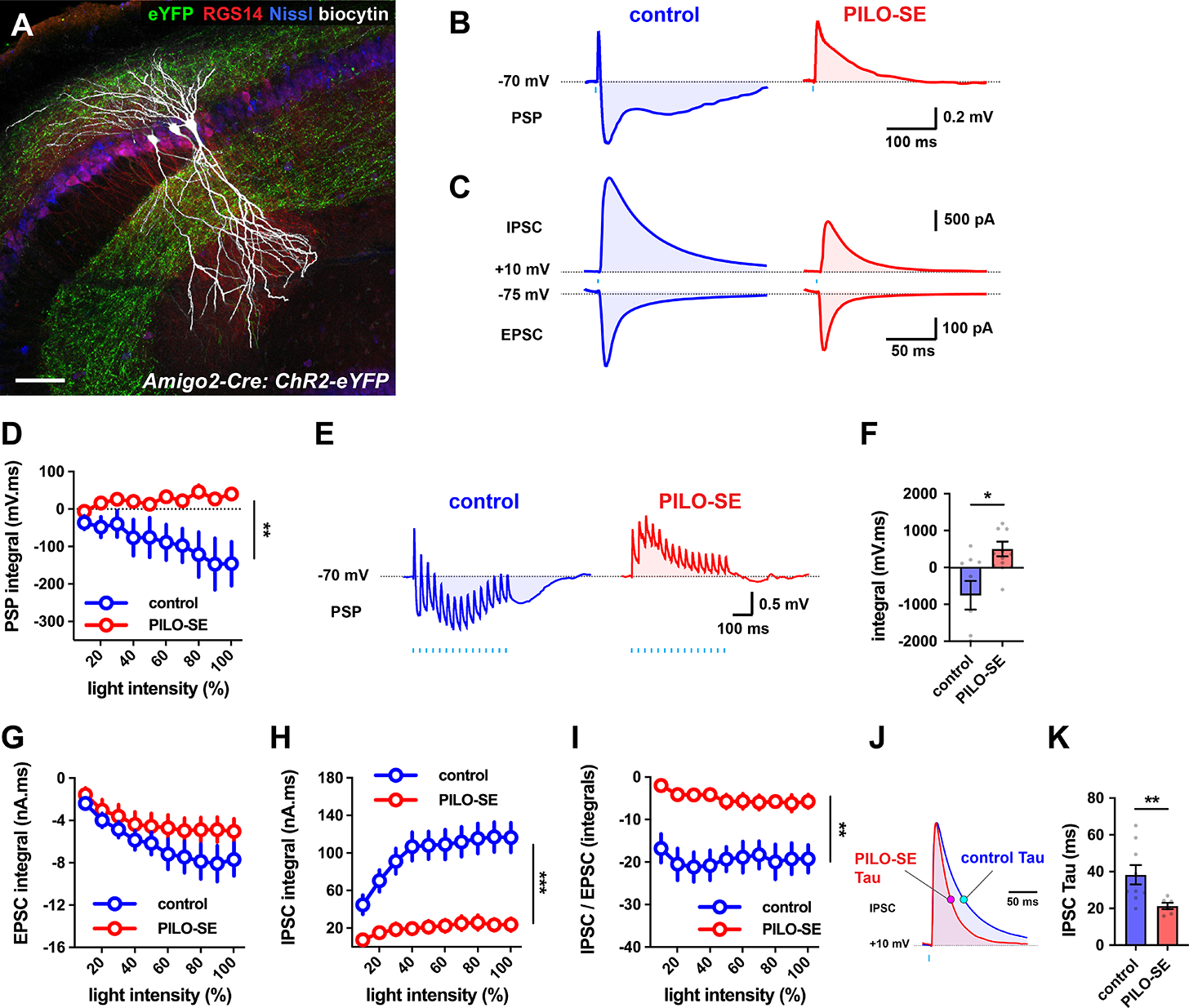
Figure 3. The inhibitory-excitatory balance of the CA2 → CA2 recurrent circuit was reduced in slices from PILO-SE.

(A) Biocytin-filled CA2 PCs (white) in a slice from intermediate hippocampus, with ChR2-eYFP-expressing CA2 PC axons (green) visible in SO and SR. CA2 PCs were labeled for RGS14 (red) and neuronal somata were visualized with Nissl stain (blue). Scale bar, 80 μm. (B) Representative averaged light-evoked PSPs from CA2 PCs from control and PILO-SE mice. (C) Representative averaged light-evoked EPSCs and IPSCs from control and PILO-SE CA2 PCs. (D) The integral of the light-evoked PSP was significantly more positive in CA2 PCs from PILO-SE mice. (E) Representative averaged PSPs evoked by 15 pulses of light delivered at 30 Hz in cells from control and PILO-SE mice. (F) The integral of the train-evoked PSP is significantly more positive in cells from PILO-SE mice. (G - I) Input-output curves of the integral of the light-evoked EPSC, the integral of the light-evoked IPSC, and the ratio of the integrals of the light-evoked IPSC and EPSC. (J) Representative averaged light-evoked IPSCs from CA2 PCs with the time constant indicated with magenta and cyan markers on the control and PILO-SE currents, respectively. (K) The time constant of the light-evoked IPSC was significantly shorter in cells from PILO-SE mice. See also Figure S2.
