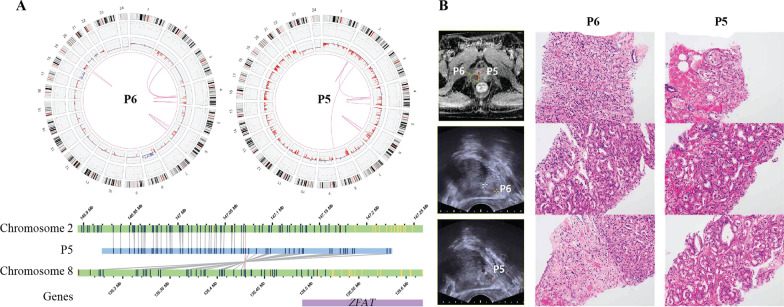
Fig. 3.
Intratumoral Heterogeneity and Complex Genomic Rearrangement. A Intratumoral genomic heterogeneity. P6 and P5 were from same patient. B MRI, US images and representative H&E staining images of P6 and P5. P5: The majority of tumor was composed of atypical tumor glands with glomeruloid feature, which is compatible with Gleason grade 4. As the second most common component, Gleason grade 3 tumor was identified. P6: On the contrary to P5 tumor, the majority of P6 tumor was comprised of atypical fused glands, showing Gleason pattern 4 (upper). Similar to P5 tumor, Gleason grade 4 tumors with glomeruloid feature was identified and admixed with Gleason grade 3 tumor, the second most common component (middle). A few scattered single tumor cells, which is compatible with Gleason grade 5 were identified as the third pattern (lower)

