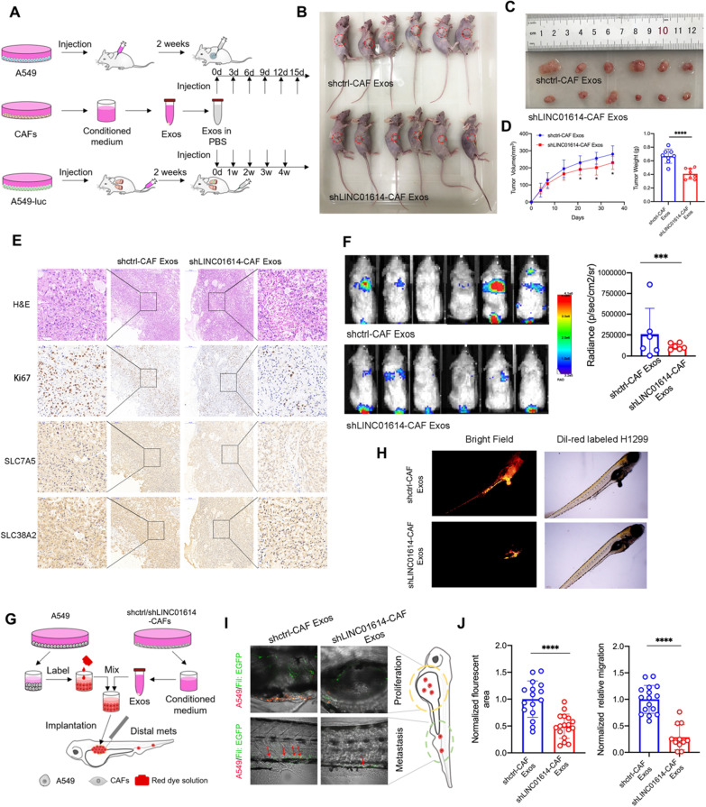
Fig. 6.
CAFs release LINC01614 in exosomes to enhance glutamine uptake and progression of LUAD in vivo. A Schematic diagram of xenograft and subcutaneous tumorigenicity in mice. B–E, Nude mice were subcutaneously xenografted with A549 cells (1 × 10⁷ cells) and treated intratumorally with CAF-derived exosomes (0.5 μg kg−1) every 3 d for 2 weeks (n = 6 mice per group). B Images of tumor engraftment in nude mice. C, D Tumor weight and growth curves. E Representative H&E staining and IHC staining for Ki67, SLC38A2, and SLC7A5. F NCG (NOD prkdc−/−IL-2Rg−/−) mice were xenografted with A549-luc cells (5 × 10⁶ cells) through tail vein injection and treated intravenously with CAF-derived exosomes once a week for 4 weeks (n = 6 mice per group). Representative bioluminescent images for lung metastasis. Scanning was performed 8 weeks after tumor implantation. G Schematic diagram of implantation of cancer cells in zebrafish embryos (Fli1:EGFR). A549 cells were labeled with Dil implanted into the perivitelline space of each zebrafish. After 24 h, the exosomes (5 ng) isolated from CAFs transduced without (shctrl) or with shRNA for LINC01614 (shLINC01614) were injected into the (Fli1:EGFR) zebrafish embryos. Dissemination of A549 was monitored. H Representative images of Dil-red-labeled A549 cells in zebrafish embryos with indicated treatments. I Representative confocal microscopy images of the dissemination of implanted DiD-labeled A549 cells in zebrafish embryos (Fli1:EGFP) injected with CAF-derived exosomes (5 ng) (n = 15). The approach schema is illustrated. J Quantification of total numbers of disseminated and metastatic cells in the primary tumor surroundings (upper) and the trunk regions (lower) of zebrafish. For D, J means ± s.d. are shown, and independent sample t-tests determined P values. *P < 0.05, **P < 0.01, ***P < 0.001. Exos exosomes, LUAD lung adenocarcinoma

