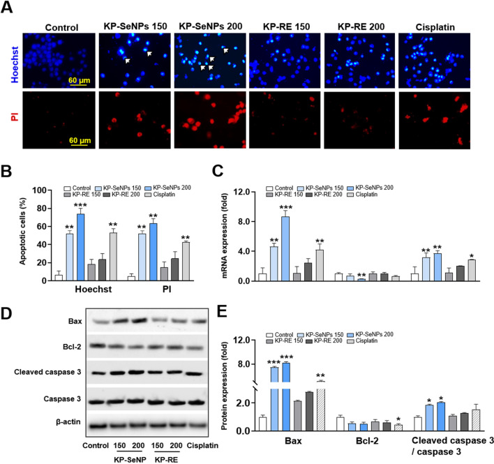
Fig. 4.
Apoptosis induced by KP-SeNPs in AGS cells. A Hoechst staining and propidium iodide (PI) staining images of AGS cells treated with KP-SeNPs (150, 200 µg/mL) and KP-RE (150, 200 µg/mL). Cisplatin (50 µM) was used as the positive control. The white arrows indicate fragmented nuclei with condensed chromatin. B Images were analyzed through the Image J software. C Effect of KP-SeNPs and KP-RE on the regulation of apoptosis-related gene expression in AGS cells. Cisplatin (50 µM) was used as the positive control. D Apoptosis-related protein expression visualized by western blotting. E Western blots were analyzed through Image J software. *p < 0.05, ** p < 0.01, ***p < 0.001 compared with control

