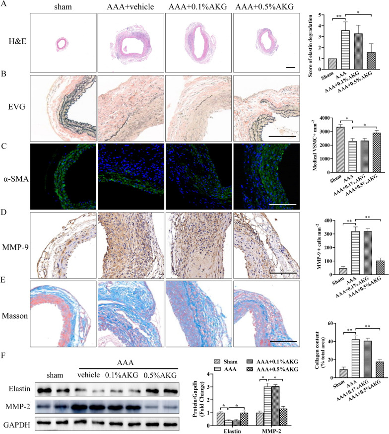
Fig. 2.
Role of AKG in elastic fibers, ECM decomposition and vascular remodeling. A Typical images showing HE staining for aortic cross-sections (n = 6). Bars = 200 μm. B Typical EVG staining as well as elastin decomposition quantification within aortic sections of specific mouse groups (n = 6). Bars = 50 μm. C Representative image and quantification of immunostaining for α-SMA (green). Nuclei were stained with DAPI. (n = 6). Bars = 50 µm. D Typical images as well as IHC staining for MMP-9 in the indicated groups (n = 6). Bars = 50 µm. E Representative image and quantification of Masson trichrome staining (n = 6). Bars = 50 µm. F The active-MMP2 (68 kDa) and elastin expression were analyzed by western blot (n = 4).*P < 0.05, **P < 0.01

