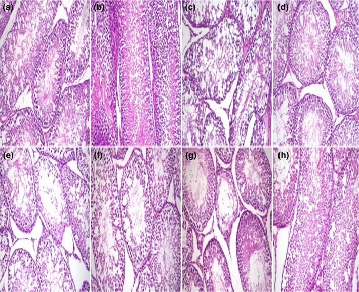
FIGURE 3.

Histopathology of rat testis. (a) Well‐organized seminiferous tubules with normal germinal epithelium in G1 (control) and (b) G2 (BM group). (c) Moderate diffuse degeneration, decrease in the thickness of lining epithelium, and vacuolation of Sertoli cells in G3 (aflatoxin group). (d) Mild degeneration of seminiferous tubules in G4 (aflatoxin‐BM group). (e) Thinning of the lining epithelium and desquamated spermatocytes and early spermatids in the lumen in G5, (f) partially alleviation of testicular lesions in G6, (g) severe diffuse degeneration in the seminiferous tubules and intraluminal infiltration of homogenous hyalinized eosinophilic material in G7 (STZ‐aflatoxin), (h) mild degeneration of seminiferous tubules in G8 (STZ‐aflatoxin and BM groups). H and E stain ×200
