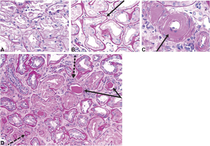
Fig. 2.
Histology images showing tubulointerstitial changes seen in diabetic kidney disease. A Normal kidney cortex. B Thickened tubular basement membrane and interstitial widening. C Arteriole with an intimal accumulation of hyaline material with significant luminal compromise. D Renal tubules and interstitium in advancing diabetic kidney disease, with thickening and wrinkled tubular basement membranes (solid arrows), atrophic tubules (dashed arrow), some containing casts, and interstitial widening with fibrosis and inflammatory cells (dotted arrow). All sections stained with period acid-Schiff stain, original magnification ×200. Reprinted with permission from American Society of Nephrology (Alicic et al. [11])

