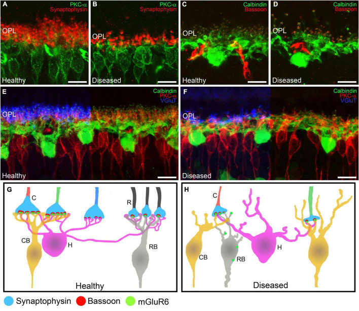
FIGURE 5.
Alterations in retinal cell connectivity in IRDs. (A–F) Retinal cross-sections immunostained with different retinal cell markers in a healthy mouse retina and in a retina from an IRD model. (A,B) Shows the dendrites of ON rod bipolar cells (PKCα, green) making contacts with photoreceptor axon terminals (synaptophysin, red). (A) Average synaptic contact density and morphology of rod bipolar cell dendrites in the OPL in healthy retinas. (B) Degeneration of ON rod bipolar cell dendrites and loss of photoreceptor synaptic terminals in IRDs retinas. (C,D) Horizontal cells (calbindin, green) being part of the ribbon synapse (Bassoon, red) along with photoreceptors. (C) Average synaptic contact density and morphology of horizontal cell dendrites in healthy retinas. (D) Loss of horizontal cell dendrites and synaptic ribbons from photoreceptors in IRDs. (E,F) Horizontal cells (calbindin, green) and ON rod bipolar cells (PKCα, red) making synaptic contacts with axonic terminals from photoreceptors (VGluT, blue). (E) Average synaptic contact density and morphology of horizontal and ON rod bipolar cell dendrites in healthy retinas. (F) Dendritic loss of horizontal and ON rod bipolar cells and degeneration of synaptic connections with photoreceptor axonic terminals. (G,H) Schematic depiction of the normal synaptic connections from rods and cones to bipolar cells in the OPL. Synaptophysin is located in the presynaptic terminals from rods and cones. Bassoon is part of the synaptic ribbon and mGluR6 is localized in the synaptic tips of the bipolar cell dendrites. During synaptic remodeling induced by IRDs (B) there are alterations in the synaptic contacts from the OPL. Rod bipolar cells make contacts with cone terminals and mGluR6 is shown delocalized around the somas of bipolar cells. OPL: outer plexiform layer; C: cone; R: rod; CB: cone bipolar cell; H: horizontal cell; RB: rod bipolar cell. Scale bars: 10 μm.

