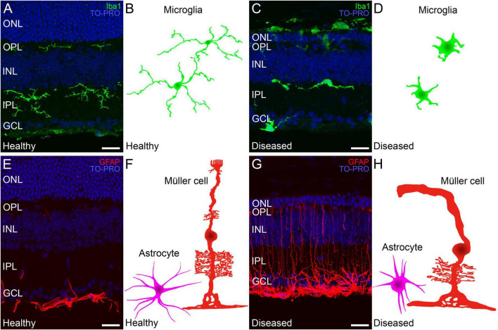
FIGURE 6.
Changes in Müller cells and microglia during retinal degeneration. Retinal cross-sections immunostained with different retinal glial cell markers followed by a schematic drawing of each cell in a healthy mouse retina and in a retina from an IRD model. (A,C) Microglia immunostained against Iba1 (in green) and TO-PRO 3 iodide-stained nuclei (in blue). (A) In healthy retinas microglial morphology is ramified and cells are localized in the IPL, OPL and GCL. (C) Ameboid morphology from active microglia in a retina with an IRD. (B,D) Schematic depiction of the morphology of microglial cells in healthy (B) and diseased conditions (D). (E) In healthy retinas GFAP (red) marker selectively immunostains astrocytes in the GCL. (G) In IRDs, astrocytes display a hypertrophied morphology and Müller cell activation causes increased GFAP immunoreactivity throughout the cell length. (F,H) Schematic depictions of representative Müller cells and astrocytes in healthy conditions (F) and affected by retinal degeneration (H). ONL: outer nuclear layer; OPL: outer plexiform layer; INL: inner nuclear layer; IPL: inner plexiform layer; GCL: ganglion cell layer. Scale bars: 20 μm.

