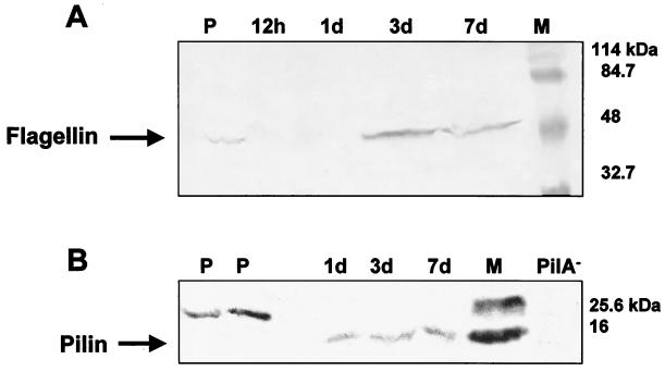
FIG. 4.
Immunoblot of b-type flagella (A) and type IV pili (B) of whole P. putida cells grown in minimal medium in a chemostat or attached to silicone surface during biofilm development. Whole cells were analyzed by SDS-PAGE, and the proteins were electroblotted onto nitrocellulose membranes (3). The membranes were probed with polyclonal b-type flagella antibodies (A) or monoclonal type IV pilus antibodies (B). Goat anti-rabbit immunoglobulin G conjugated to horseradish peroxidase was used as the secondary antibody. Antibody binding was detected by colorimetric analysis (3). M, marker; PilA−, type-IV-pilus-deficient P. aeruginosa PA416 (49); P, planktonic, chemostat-grown P. putida cells; 12 h and 1 d, attached P. putida cells after 12 h and 1 day of attachment time, respectively.

