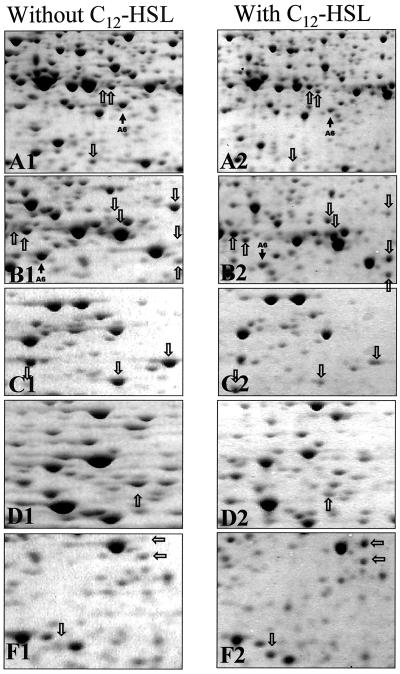
FIG. 5.
Enlarged partial 2-D gels of crude protein extracts of P. putida in the absence (A1 to F1) and presence (A2 to F2) of the 3OC12-HSL signal molecule. The sections A1 to F1 and A2 to F2 correspond to the boxes A to F shown in Fig. 2. Arrows indicate differences in the 2-D protein pattern of chemostat-grown cells in the absence and presence (10 μM) of the C12-HSL signal molecule.

