FIGURE 2.

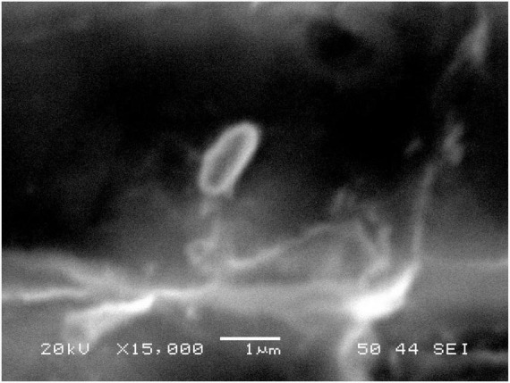
Scanning Electron Microscopy of Isolated Bacillus Licheniformis. The photography was taken by a JEOL JSM-6360LV using a magnification of 15,000X and an accelerating voltage of 20 kV.

Scanning Electron Microscopy of Isolated Bacillus Licheniformis. The photography was taken by a JEOL JSM-6360LV using a magnification of 15,000X and an accelerating voltage of 20 kV.