
FIGURE 2.
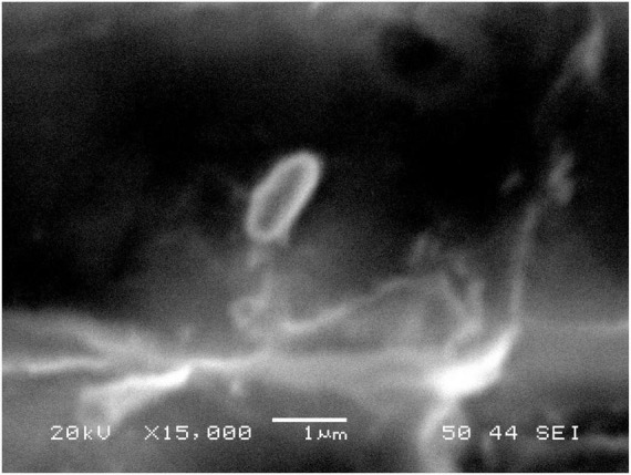
Scanning Electron Microscopy of Isolated Bacillus Licheniformis. The photography was taken by a JEOL JSM-6360LV using a magnification of 15,000X and an accelerating voltage of 20 kV.
Official websites use .gov
A
.gov website belongs to an official
government organization in the United States.
Secure .gov websites use HTTPS
A lock (
) or https:// means you've safely
connected to the .gov website. Share sensitive
information only on official, secure websites.
Scanning Electron Microscopy of Isolated Bacillus Licheniformis. The photography was taken by a JEOL JSM-6360LV using a magnification of 15,000X and an accelerating voltage of 20 kV.