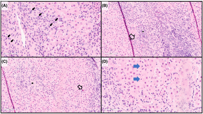
FIGURE 1.

Histology of AIH (hematoxylin and eosin stains). (A) the inflammatory infiltrate often includes abundant and conspicuous plasma cells (arrows). (B) Interface hepatitis is characteristic, with the inflammatory infiltrate extending beyond the limiting plate into the lobule (arrow), including abundant and clustered plasma cells (star). (C) Lobular inflammation (star) with scattered acidophil bodies (necrotic hepatocytes) may also be present (arrow). (D) Emperipolesis of inflammatory cells by hepatocytes may be observed (arrows)
