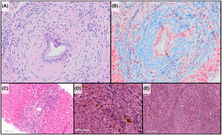
FIGURE 2.

Histology of PSC. (A) Characteristic periductal fibrosis is seen in portal tracts (hematoxylin and eosin stains). (B) Masson trichrome stain highlights the dense collagen fibrosis surrounding the residual ductal epithelium. (C) Ductular reaction/proliferation is seen, likely as a result of downstream biliary obstruction. (D) Longstanding PSC leads to a cholestatic pattern of cirrhosis with inspissated bile, extravasated bile, and canalicular bile all be seen in this image. (E) Cirrhotic nodules, in an explanted liver of a child with history of PSC
