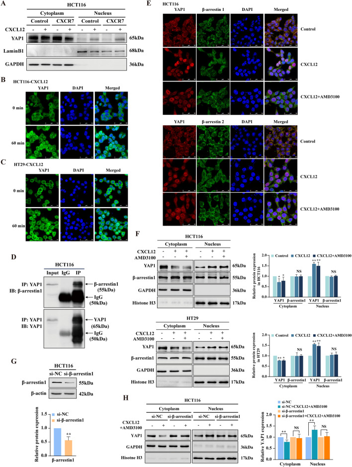
Fig. 4.
CXCR7/β-arr1-mediated biased signal induces YAP1 nuclear translocation in CRC cells. A Western blot analysis of YAP1 expression in cytoplasmic and nuclear extracts of HCT116Control and HCT116LV−CXCR7 cells treated with or without CXCL12 (100 ng/ml) for 60 min. GAPDH and Lamin B1 were used as cytoplasmic and nuclear loading control, respectively. B, C YAP1 localization evaluated by immunofluorescence (IF) in HCT116 and HT29 cells treated with or without CXCL12 (100 ng/ml). YAP1 was labeled with Alexa Fluor® 488 donkey anti-rabbit secondary antibodies, nuclei were visualized with DAPI, shown in blue. Scale bars, 50 µm. D Analysis of endogenous YAP1-β-arr1 interaction in HCT116 cells by Co-immunoprecipitation (Co-IP). Normal rabbit IgG antibodies were used as control. E IF staining was performed to determine the colocalization of YAP1 (red) and β-arrestin1 (green) or β-arrestin2 (green) in HCT116 cells treated with CXCL12 (100 ng/ml) in the presence of AMD3100 (2 μM). DAPI was used for nuclear staining. Scale bars, 50 µm. F Western blot analysis of YAP1 and β-arrestin1 expression in cytoplasmic and nuclear extracts of HCT116 and HT29 cells treated with or without CXCL12 (100 ng/ml) in the presence of AMD3100 (2 μM). GAPDH and Histone H3 were used as cytoplasmic and nuclear loading control, respectively. G Western blot analysis of β-arrestin1 expression in HCT116 cells transfected with β-arrestin1 siRNA. β-actin was used as loading control. H Western blot analysis of YAP1 expression in cytoplasmic and nuclear extracts of HCT116 cells transfected with β-arrestin1 siRNA or siNC and treated with or without CXCL12 (100 ng/ml) plus AMD3100 (2 μM). GAPDH and Histone H3 were used as cytoplasmic and nuclear loading control, respectively. Bars are means ± SD; *P < 0.05, **P < 0.01, ***P < 0.001, NS stands for no significance (n = 3)

