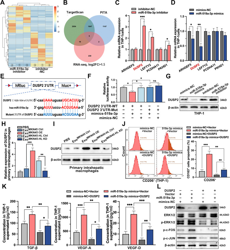
Fig. 6.
DUSP2-MAPK/ERK axis is the functional target of exo-miR-519a-3p in macrophages. A. mRNA-seq was performed in PMA-treated THP-1 cells with or without miR-519a-3p inhibitor. The heatmap showed the top 100 genes with significant differences in expression after knockdown of miR-519a-3p. B. Five mRNAs were identified that met the criteria to be upregulated (log2FC ≥ 1.5) based on mRNA-seq data and predicted to be miR-519a-3p targets based on TargetScan and PITA. C, D. The relative expression of the five targets in PMA-treated THP-1 cells transfected with miR-519a-3p NC/mimics or miR-519a-3p NC/inhibitors was detected by qRT-PCR. E. Schematic representation of the wild-type and mutant-type binding site between the 3'UTR of DUSP2 and miR-519a-3p. F. Relative luciferase activity of 3'UTR-DUSP2-luc constructs in HEK293T cells after transfection of miR-519a-3p mimics/NC. G. Expression of DUSP2 in PMA-treated THP-1 cells in which miR-519a-3p was overexpressed or knocked down was detected by Western blot. H, I. Expression of DUSP2 in primary liver macrophages from miR-519a-3p-deficient or enriched exosome-educated mice was detected by qRT-PCR and Western blot. J. Expression of CD206 (M2 marker) in miR-519a-3p overexpressing or miR-519a-3p/DUSP2 co-expressing PMA-treated THP-1 cells was determined by flow cytometry. K. Secretion of TGF-β, VEGFA, and VEGFD in miR-519a-3p overexpressing or miR-519a-3p/DUSP2 co-expressing PMA-treated THP-1 cells was detected by ELISA. L. Changes in the expression levels of the downstream cytokines p-ERK1/2, p–c-FOS, and p–c-JUN of the MAPK pathway was detected by Western blot in PMA-treated THP-1 cells transfected with miR-519a-3p mimics and DUSP2 overexpression vectors alone or together. Data are shown as mean ± standard deviation of 3 independent experiments, and statistical significance was determined using Student’s t test and one-way ANOVA test (*P < 0.05, **P < 0.01, ***P < 0.001)

