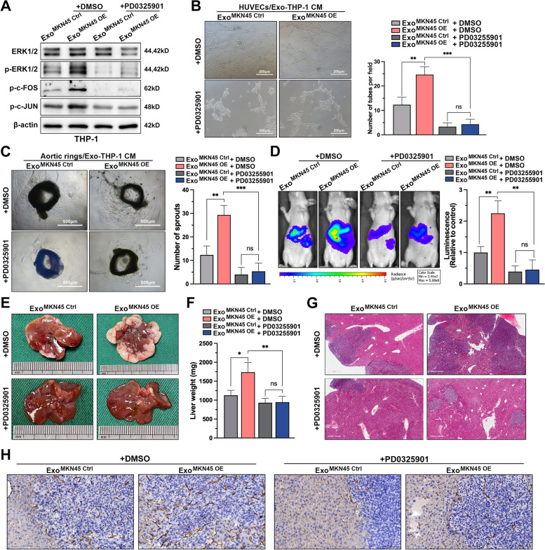
Fig. 7.
DUSP2-MAPK/ERK axis is the functional target of exo-miR-519a-3p in macrophages. A. Expression levels of downstream cytokines p-ERK1/2, p–c-FOS, and p–c-JUN in the MAPK pathway after addition of MEK1 inhibitor (PD0325901) to PMA-treated THP-1 cells pre-incubated with control or exosomes overexpressing miR-519a-3p. B. Tubule formation assays of HUVECs were performed after 48 h of incubation with conditioned medium (with or without MEK1 inhibitor) from PMA-treated THP-1 cells pre-incubated with exo-miR-519a-3p. Scale bar = 200 μm. C. Effect of conditioned medium (with or without PD0325901) of PMA-treated THP-1 cells pre-incubated with miR-519a-3p-riched or control exosomes on vascular sprouting in mouse aortic rings. Scale bar = 500 μm. D. Mice educated with miR-519a-3p-overexpressed or controlled exosomes were simultaneously treated with PD0325901 (20 mg/kg, gastric lavage every 2 days) or DMSO followed by portal vein injection with luciferase-labeled MKN45 cells. The fluorescence signal in the metastases was detected and quantified by IVIS. E–H. The mice were then sacrificed, and the livers were harvested for weighing, photography, H&E and CD31 staining. Scale bar = 500 μm. Data are shown as mean ± standard deviation of 3 independent experiments, and statistical significance was determined using one-way ANOVA test (*P < 0.05, **P < 0.01, ***P < 0.001)

