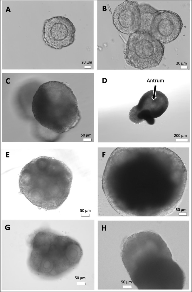
Fig. 1.
A Preantral follicle with 2–3 layers of granulosa cells collected after collagenase digestion of ovary (B) Individually isolated follicles seeded in HA gel on day 2 of culture. Follicles tended to aggregate during gelling process. C Day 4 of culture. Follicles growing in different planes within HA gel (D) HA-encapsulated on day 9 of culture. Antrum formation discernible (E, F) FL-cluster from fresh ovary embedded in HA gel shown at start of culture and on day 6. Granulosa cell proliferation evident and FL-cluster takes on an”organoid” appearance (G, H) FL-cluster mechanically isolated from vitrified ovary shown on day 2 and day 8 of culture

