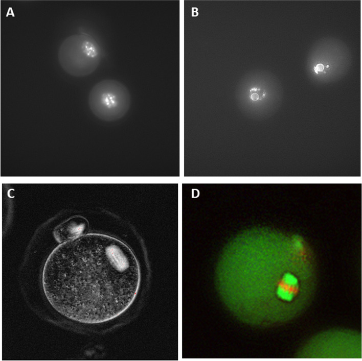
Fig. 2.
A Chromatin staining at outset of culture showing GV oocyte with non-surrounded chromatin staining (NSN) pattern (B) oocyte from growing follicle at time of antrum formation on day 9. GV oocyte with SN staining pattern. Chromatin condensed and forming ring around the nucleolus (C) Live imaging of metaphase II oocyte and spindle using polarized light and birefringence (D) Immunofluorescent staining of metaphase II oocyte showing normal meiotic spindle with fluorescent microtubules running to poles and chromosomes aligned on equatorial plate. 400 × magnification

