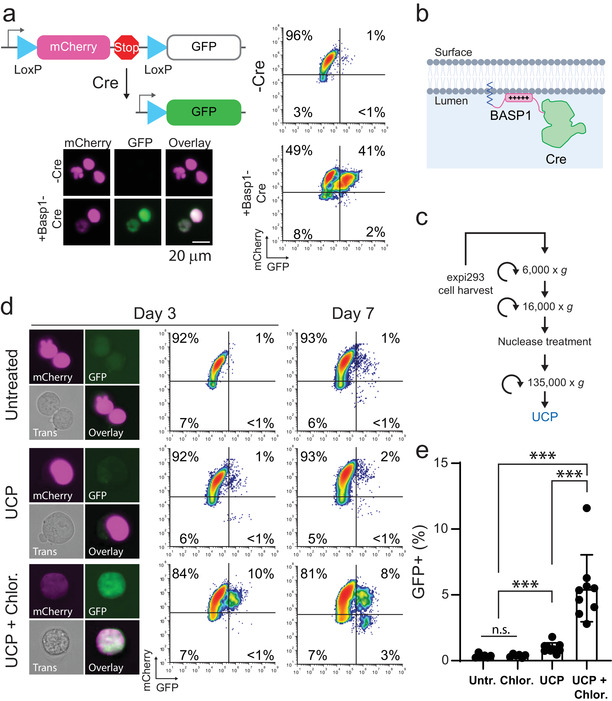
FIGURE 1.

Ultracentrifuge pellets contain Cre activity that is enhanced by an endosome escaping agent. (A) Schematic of Cre “stoplight” reporter system. GFP is expressed only after Cre‐mediated excision of the mCherry and Stop codon. Flow cytometry and microscopy of stable reporter cells either untreated or transiently transfected with BASP1‐Cre plasmid. Scale Bar = 20 μm. (B) Schematic of Cre protein fused to Basp1 N‐terminal peptide (amino acids 1–10) for localization to the EV lumen. (C) Schematic of EV purification procedure. (E) Representative images and flow cytometry plots of reporter cells 3 and 7 days after addition of UCP isolated from cells transfected with Expifectamine +/‐ 50 μM chloroquine. Plot axes are the same as in (A). (E) Bar graph showing the percentage of GFP+ reporter cells on day 3 from nine independent experiments (N = 9, ***P < 0.001, n.s. not significant: P > 0.05, Mann‐Whitney U‐test)
