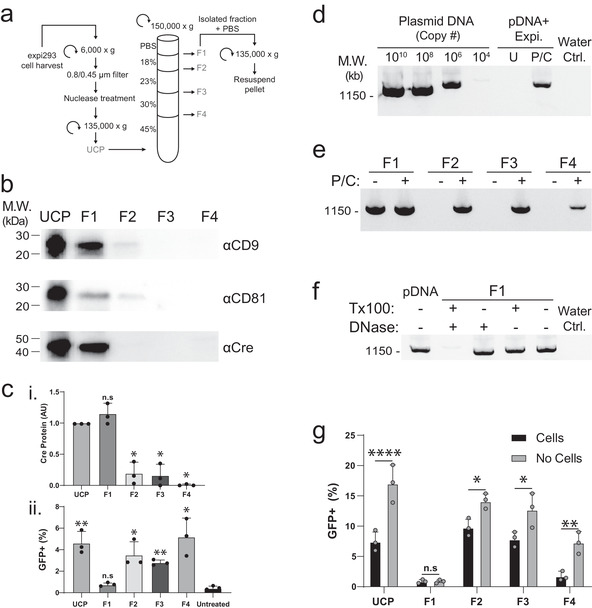
FIGURE 2.

Cre activity present in UCP is biochemically separable from Cre protein and EVs. (A) Schematic of differential centrifugation and iodixanol density gradient used to separate UCP isolated from cells transfected with Expifectamine into four fractions (F1‐4). (B) Western blot analysis of UCP and density gradient fractions with antibodies against Cre and the EV markers CD9 and CD81. Equal particle numbers (1e10 particles) were loaded in each lane. (C) Quantification of Cre protein by Western blot densitometry (i); statistical comparison of each density gradient fraction relative to UCP (*, P < 0.0005, n.s., not significant, Mann‐Whitney U‐test). (ii) Quantification of GFP+ reporter cells 3 days after treatment with 50 μM chloroquine and either UCP or F1‐4 analysed by flow cytometry; statistical comparison of each sample relative to untreated controls. (*, P < 0.05; **, P < 0.005; n.s., not significant, P > 0.05; unpaired Mann‐Whitney U test). (D‐F) Agarose gels stained with SYBR safe DNA dye showing PCR amplification from the indicated samples. (D) Control plasmid (lanes 1–4); lanes 5–6: plasmid complexed with Expifectamine reagent, treated with nuclease, followed by phenol chloroform extraction (P/C) or column purification (U) prior to PCR amplification. Water Ctrl. is a no‐plasmid control containing all other PCR components. (E) Density gradient fractions F1‐4 were processed with (+) or without (‐) phenol chloroform extraction (P/C) prior to PCR amplification. (F) PCR amplification of control plasmid (pDNA) or F1 material pre‐treated with the indicated compounds. Water Ctrl. is a no‐plasmid control containing all other PCR components. (G) Quantification of GFP+ reporter cells treated with 50 μM chloroquine and either UCP or F1‐4. For the No Cells condition, a mock transfection was performed where no cells were included in the transient transfection with Cre plasmid; samples were otherwise processed identically. Cells analysed 4 days post treatment, with no media exchange for either condition (*, P < 0.05; **, P < 0.005; ****, P < 0.0001; n.s., not significant: P > 0.05; two‐way ANOVA)
