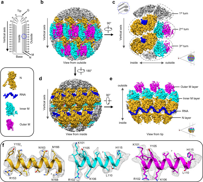
Fig. 1. Sub-particle reconstruction of VSV trunk at near-atomic resolution.
a Schematic diagram of the VSV virion. Blue circle marks the region used for sub-particle reconstruction. b, d CryoEM density maps of partial VSV nucleocapsid shown in two opposite views. Only intact N, intact M and RNA are colored for clarity; other densities are gray. N subunits are colored in goldenrod, inner M in cyan, outer M in magenta and RNA in blue (inset). c, e Cross sections of cryoEM density maps viewed from side and tip. f CryoEM density maps (mesh) of α-helices from three kinds of subunits.

