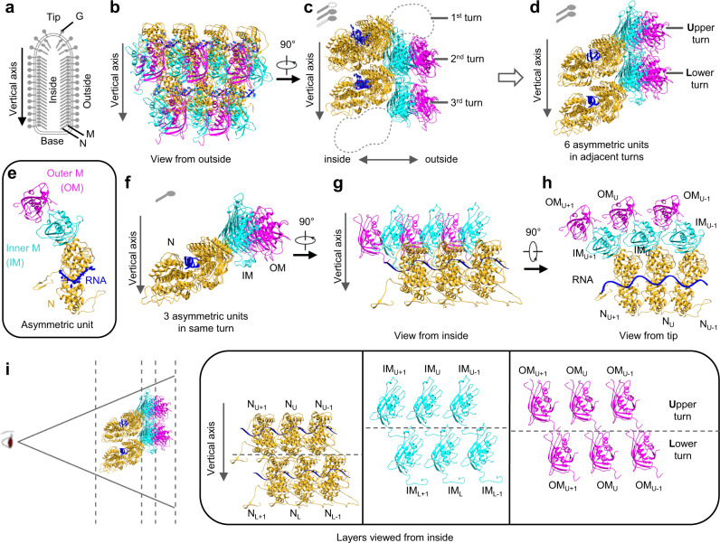
Fig. 2. Atomic model of the partial VSV nucleocapsid.
a Schematic diagram of the VSV virion. b, c Two orthogonal views of the atomic model, which contains 7 N (goldenrod), 7 inner M (cyan) and 5 outer M (magenta). Only intact N, intact M and RNA are built and colored, as in the cryoEM map. d Six asymmetric units from two turns derived from (c). e One asymmetric unit, including 1 N, 1 IM and 1 OM, together with 9 RNA nucleotides. f–h Three asymmetric units from the same turn shown in three orthometric views. i Layer expansion of (d) from the central axis of the virion. Each subunit is labeled based on its lateral relationship (+1 or −1, along the left-handed helix) and vertical position (Upper (U) and Lower (L) turns).

