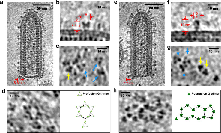
Fig. 4. In situ structures of VSV G trimers and their hexagonal/pentagonal distribution.
a A 6 Å-thick density slice from a reconstructed tomogram showing a representative VSV virion purified without a density gradient centrifugation step. The 8.5 nm-thickness of glycoproteins indicates that most G are in prefusion conformations. b, c VSV G trimers in prefusion mostly or postfusion conformation occasionally. b The side view of a representative G trimer in prefusion conformation from 4452 trimers obtained by template matching; c The top views of G trimers. Trimers in prefusion conformation, are pointed by blue arrows, while those in postfusion conformation by yellow arrows. d Example of a hexagonal tile formed by prefusion G trimers on the viral membrane. e A 6 Å-thick density slice from a reconstructed tomogram showing a representative VSV virion purified with a density gradient centrifugation step. The 12 nm-thickness of glycoproteins indicates that most G are in postfusion conformations. f, g VSV G trimers in postfusion mostly or prefusion conformation occasionally. f The side view of a representative G trimer in postfusion conformation from 6030 trimers obtained by template matching; g The top views of G trimers. Trimers in postfusion conformation, are pointed by yellow arrows, while those in prefusion conformation by blue arrows. h Two connected hexagonal tiles joined to a pentagonal tile formed by postfusion G trimers on the viral membrane.

