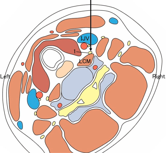
Fig. 3.

A schematic illustration of the anatomical structures in axial image (from the view of the operator standing on the cranial side of the patient) at C6 level of the cervical spine. CA, the right common carotid artery; IJV, the right internal jugular vein; VA, the right vertebral artery; LCM, the right longus colli muscle; ASM, the right anterior scalene muscle; 1, the right cervical sympathetic nerve trunk; 2, the right vagus nerve; 3, the right phrenic nerve; 4, the right brachial plexus. (This figure was constructed using an article by Akashi et al. [22].)
