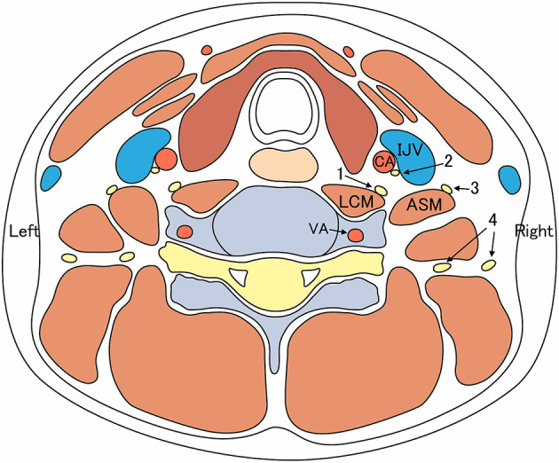
Fig. 4.

A schematic illustration of the puncture tract (shown as a large black arrow) for the right internal jugular vein, with 45-degree neck rotation to the left (the same anatomical schema as seen in Fig. 3) . If vertical puncture from anterior to posterior is performed on axial (short-axis) view, the cervical sympathetic nerve and longus colli muscle are located in the path of the puncture tract. IJV, the right internal jugular vein; LCM, the right longus colli muscle; 1, the right cervical sympathetic nerve trunk.
