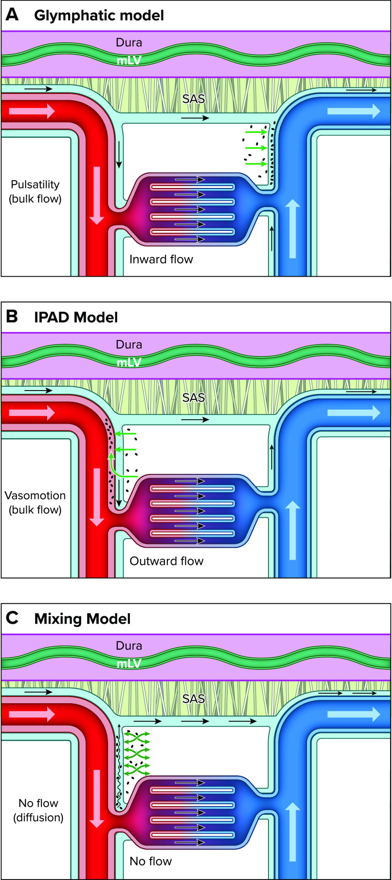
FIGURE 1.
Schematic presentation of the three models of perivascular brain waste clearance Note that in all illustrations the dura mater is represented by a purple-colored membrane with meningeal lymphatic vasculature (mLV) in shown in green. The dura mater is separated from the subarachnoid space (SAS) by the arachnoid membrane (arachnoid trabeculae shown in gray). The cerebrospinal fluid (CSF) flowing along the periarterial and perivenous channels is illustrated in light blue. The arteries and veins are shown in red and blue, respectively. A: the glymphatic system model posits that waste solutes are cleared from the brain in the following manner: CSF flows into the brain along the perivascular spaces of the arteries (i.e., inward flow) and is propelled (via bulk flow) into the interstitial fluid (ISF) compartment, which forces waste toward the perivenous conduits, from where it merges and exits via meningeal lymphatics. B: the “intramural periarterial drainage” (IPAD) model poses that CSF flows into the brain along the periarterial channels and waste solutes from the ISF exit via walls of the arteries and are transported in a direction opposite to blood flow driven by vasomotion (outward flow). C: the mixing model poses that there is no net flow (no flow) along the penetrating cerebral vasculature. Instead, waste transport from the ISF to the periarterial perivascular spaces (PVS) is facilitated by “mixing” from physiological motion (e.g., vascular pulsatility), as illustrated by the green “mixing” arrows. Furthermore, according to the mixing model, bulk flow is present only on the surface of the brain along the large pial arteries in the subarachnoid space, which creates a favorable concentration gradient for waste egress by diffusion from the ISF to the arterial PVS in the tissue and then toward the pial surface.

