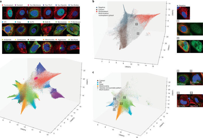
Fig. 4. Visualization of learned single-cell features.
a, Overview of all single-cell clusters (except Negative). Notably, the patterns organize into cytosolic (bottom-left) and nuclear (top-right) meta-clusters. b, Cells with both the Cytosol and the Nucleoplasm labels lie between the two respective clusters. The majority of negative cells cluster into a separate corner with a slight overlap with Cytosol and Nucleoplasm clusters. c, Cells labeled with Vesicles and punctate pattern (Ves. Punct.) in the cytosol, endoplasmic reticulum (ER) and the Golgi apparatus cluster together at the opposite end to the nuclear patterns in the three-dimensional UMAP plot. Cells labeled Vesicles can be found in these other clusters, capturing the cellular dynamics of the class. Actin fil., Actin filaments; Golgi, Golgi apparatus; Int. Fil, Intermediate filaments; M. Spindle, Mitotic spindle; Nuc. Bodies, Nuclear bodies; Nuc. Fib. C., Nucleolar fibrillar center; Nuc. Membrane, Nuclear membrane; Nuc. Speckle, Nuclear speckle; Pl. Membrane, Plasma membrane.

