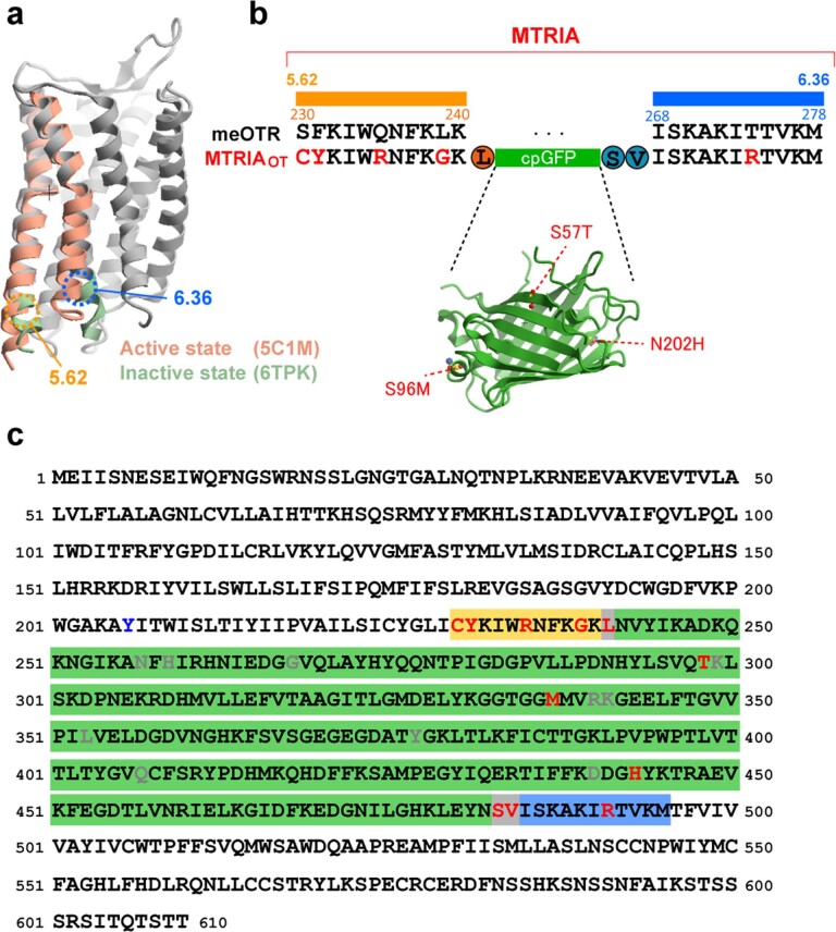
Extended Data Fig. 2. Sequence of our fluorescent OT sensor.
a, Superposition of active- and inactive-GPCR structures, adapted from the Protein Data Bank (PDB) archives (IDs: 5C1M and 6TPK). TM5–TM6 regions of active and inactive states are colored pink and green, respectively. b, Alignment of the TM5–TM6 region between meOTR and MTRIAOT; the structure of cpGFP is adapted from a PDB archive (ID: 3SG2). Mutations introduced in MTRIAOT are shown as red. c, Full amino acid sequence of MTRIAOT. Mutations in MTRIAOT, the point mutation in MTRIAOT-mut, and mutations of cpGFP that were adapted in the other fluorescent sensors are shown in red, blue, and gray, respectively.

