
FIGURE 7.
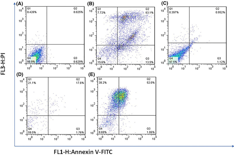
Cytometry analyses of MCF‐7 cells treated with control (A), free DOX (B), CS‐MMT‐NCQDs (C), CS‐MMT‐DOX (D), and CS‐MMT‐NCQDs‐DOX (E). In cases where non‐gated cells in FL1 (Annexin V) versus FL3 (P.I.) channels were used, the fluorescence intensity of 5000 events was measured. The lower left quadrant cells are defined as viable, the lower right quadrant cells as apoptotic, the upper left as necrotic, and the upper right quadrant cells as late apoptotic.