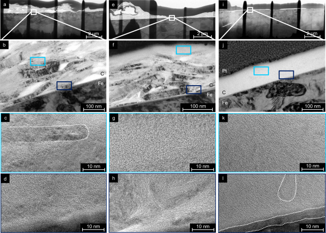
Fig. 4. Bright field transmission electron microscopy (BF-TEM, upper half) and high-resolution transmission electron microscopy (HR-TEM, lower half) images.
Taken from TEM lamella prepared in an unworn region (a–d), after sliding under 50 MPa and in ≤5% RH humidity (e–h), and after sliding under 1 GPa and in 24% RH humidity (i–l). Marked in b, f, and j are the iron substrate (Fe), the graphite layer (C), as well as the protective platinum layer (Pt) on the TEM lamella. Marked in light and dark blue are the approximate positions of the HR-TEM studies in the BF-TEM images.

