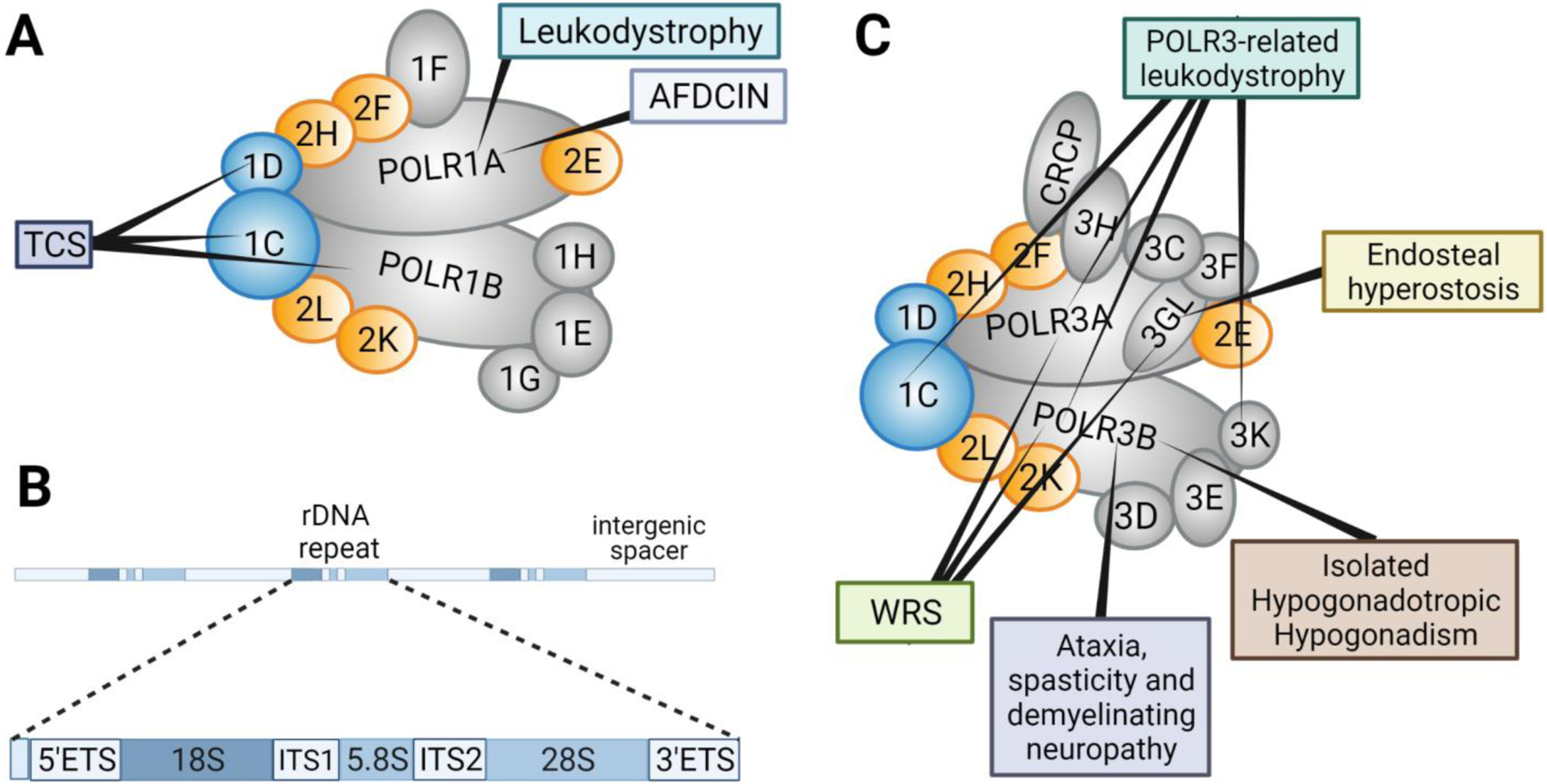
Figure 1.

RNA Polymerase I subunits, rDNA repeat, and RNA Polymerase III subunits. A) Schematic representation of Pol I in humans. Shared Pol I and III subunits POLR1C and POLR1D are in blue and subunits shared across Pols I, II, and III are in orange. B) Structure of the rDNA repeat. The promoter elements are denoted in light blue. The 47S transcript consists of the 5’ Externally Transcribed Spacer (ETS), 18S rRNA, Internally Transcribed Spacer (ITS)1, 5.8S rRNA, ITS2, 28S rRNA, and the 3’ETS. These rDNA repeats are separated by intergenic spacers (white). C) Schematic representation of Pol III subunits in humans. Pol III isoform with subunit POLR3GL is represented, but it should be noted that an alternative form of Pol III exists with subunit POLR3G (not shown). Shared subunits are indicated as in A). Disorders associated with Pol I and III subunits are indicated in boxes.
