
Table 2.
MDP derivatives with cytotoxic /antitumor activity.
| CHEMICAL STRUCTURE | BIOLOGICAL ACTIVITY | REF. | |
|---|---|---|---|
| 1. |
L-mifamurtide
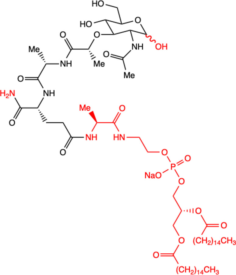
Conjugation of MDP-L-alanine to the phospholipid dipalmitoyl phosphatidylethanolamine |
Inhibition of lung metastasis dissemination and development, slowing tumor growth in mice model.
Alone has no effect on osteosarcoma cells proliferation: MOS-J and KHOS in vitro. With Zoledronic Acid combination significantly inhibited primary osteosarcoma progression. Human KHOS and murine K7M2 osteosarcoma cell lines; syngeneic (MOS-J) and xenogeneic (KHOS) mice model of osteosarcoma |
(18) |
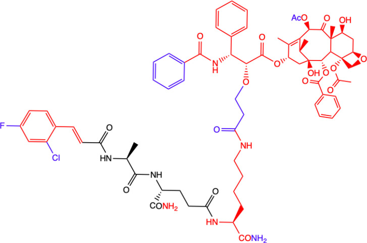
| 2. |
MTC-220

Full structure of paclitaxel (PTX) combined with MDP analogue – MDA; 4-Cl substituted ring |
Inhibition of tumor growth and metastasis in transplanted Lewis lung carcinoma (LLC) cells and breast cancer 4T1 cells mouse model.
Suppressed Myeloid Derived Suppressor Cells (MDSCs) accumulation in the spleen and bone marrow. Suppresses inflammatory cytokines in tumor tissue. Human breast (MDA-MB-231, MCF-7), ovarian (A2780, ES-2), and lung (H460, A549, H1975) tumor cell lines; 4T1 mammary carcinoma; in vitro and in C57BL/6 mice |
(71) |
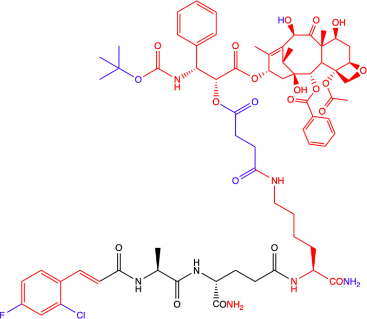
| 3. |
DY-16-43

Full structure of PTX combined with; 4-Cl substituted ring |
Sensitizes to PTX therapy and prevented metastasis in LLC mouse model – represents a potential adjunctive therapy for the treatment of NSCLC.
HEK-Blue hNOD2-secreted alkaline phosphatase (SEAP) reporter cell line; human peripheral blood mononuclear cell (PBMC)-derived macrophages; Lewis lung carcinoma (LLC) tumor-bearing mice |
(72) |
| 4. |
Compound nr 23
Full structure of paclitaxel (PTX) combined with MDA |
This compound, like DY-16-43 showed the highest level of antagonistic activity via MDP activating NOD2 signaling but has poorer solubility under the conditions tested.
HEK-Blue hNOD2 cells; Lewis lung carcinoma (LLC) tumor-bearing mice. |
|
| 5. |
Compound nr 24

7-OH substituted 10-OAc-10-DAB, without side chain of PTX; MDA - 4-Cl and 2-F substituted ring |
The experiment showed that the full structure of PTX is necessary to maintain antagonistic activity via MDP activating NOD2 signaling, which results in sensitization of a response to chemotherapy in treating non-small-cell lung cancer (NSCLC).
Compounds labelled 24 to 28 are devoid of paclitaxel structure which resulted in significantly lower inhibitory percentages than compound DY-16-43 and nr 23. HEK-Blue hNOD2 cells; Lewis lung carcinoma (LLC) tumor-bearing mice. |
(72) |
| 6. |
Compound nr 25

10-OH substituted 10-DAB, without side chain of PTX; MDA - 4-Cl and 2-F substituted ring |
||
| 7. |
Compound nr 26

13-OH substituted 10-OAc-10-DAB, without side chain of PTX; MDA - 4-Cl and 2-F substituted ring |
||
| 8. |
Compound nr 27

Side chain of PTX with Me group, without 10-DAB; MDA - 4-Cl and 2-F substituted ring |
||
| 9. |
Compound nr 28

Side chain of PTX with nBu group, without 10-DAB, MDA - 4-Cl and 2-F substituted ring |
||
| 10. |
MDC-405
Docetaxel (PTX analogue) combined with MDA: with CONH2 group |
Excellent pharmacological profile against 4T1 breast tumor growth in mice and against metastasis.
Unsatisfactory physicochemical properties - primarily very poor water solubility. 4T1 mammary carcinoma cells |
(73) |
| 11. |
Salutaxel

Docetaxel (PTX analogue) combined with MDA: with COOH group |
High anticancer efficacy together with satisfactory pharmacological properties.
Inhibition of the growth of several tumor types in vivo. Better anticancer properties than docetaxel. Human tumor cell line xenograft models: MDA-MB-231 (breast), H1975 (lung), HCT116 (colon), and A549/T (lung for paclitaxel resistance); 4T1 mammary carcinoma cells |
(73) |
| 12. |
Compound 8f

MDP derivative combined with N6-(2-aminoethyl)adenosine |
Did not show significantly higher antiproliferative activity compared to MDP and nor-MDP but highest index of selectivity.
Lymphoid cell lines and activated peripheral blood mononuclear cells (PBMC) as in vitro model Two models of leukemia were used: the human T lymphocyte-based Jurkat cell line and a mouse lymphocytic leukemia L1210. |
(14) |
| 13. |
Compound 8g
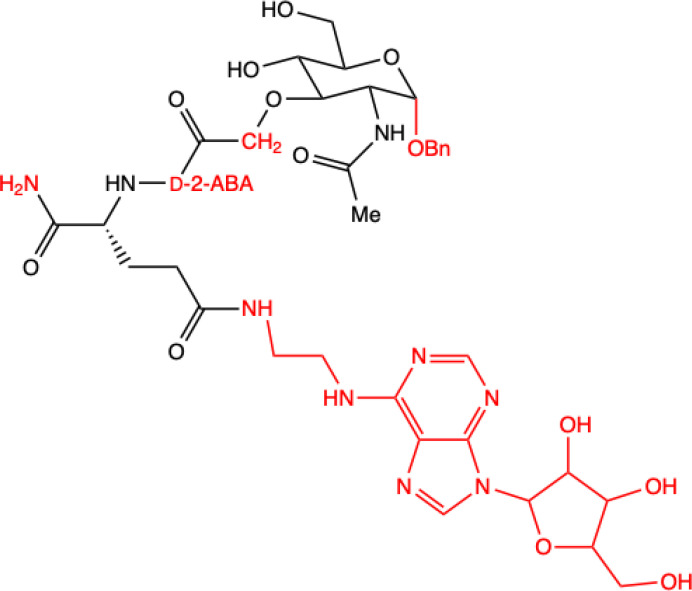
MDP derivative with abscisic acid (D-2-ABA) combined with N6-(2-aminoethyl)adenosine |
||
Green, research model/cell lines; blue, sites of change in molecular structures of paclitaxel (PTX) conjugate and MDP analogue; red, changes in structure relative to the MDP.