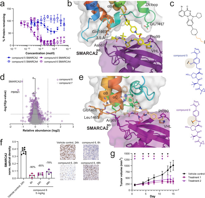
Fig. 2. Ternary structure-guided design of an in vivo active dual SMARCA2/4 degrader.
a SMARCA2 and SMARCA4 protein levels (continuous vs. dashed lines) measured by capillary electrophoresis normalised to DMSO control following compound treatment of RKO cells for 18 h (mean and standard deviation of n = 6 independent experiments for compound 6 SMARCA2, n = 5 for compound 6 SMARCA4 (both purple), n = 3 for compound 5 SMARCA2 and SMARCA4 (blue)). Values indicated by x are excluded from curve fit (hook effect). b Ternary X-ray crystal structure for VCB: compound 5: SMARCA2BD (PDB: 7Z6L, purple = VHL, yellow = 5, rainbow = SMARCA2BD). c Structures of compound 5 (blue) and compound 6 (purple) highlighting the VHL exit vector switch. d Effects of compound 6 (purple) and negative control compound 7 (grey, cis-hydroxyproline analogue of compound 6, which abrogates binding to VHL) on the proteome of NCI-H1568 cells treated with the compounds at 100 nM for 4 h. Data are plotted as the log2 of the normalised fold change in abundance against –log10 of the p value per protein from n = 3 independent experiments (two-tailed t-tests assuming equal variances). e Ternary X-ray crystal structure for VCB: compound 6: SMARCA2BD (PDB: 7Z77, purple = VHL, pale orange = 6, rainbow = SMARCA2BD). f NCI-H1568 tumour bearing mice (average tumour size ~260 mm3) were treated subcutaneously with 5 mg/kg compound 6 (n = 4 animals per time point) or vehicle control (n = 8 animals, 24 h only) and tumours were collected 6, 24 and 48 h after treatment (shades of purple). SMARCA2 levels were determined by IHC staining (representative images are shown, scale bar = 50 µm). Each datapoint represents the background-normalised optical density (OD) within the viable tumour area of one tumour section, corresponding to one individual tumour. Mean OD and standard deviations are indicated. Percentages represent the median levels of SMARCA2 signal decrease relative to vehicle (black). g NCI-H1568 tumour bearing mice (average tumour size ~210 mm3) were treated subcutaneously with 5 mg/kg compound 6 in two treatment schedules (Treatment 1/2, see methods for details, square vs. triangle in shades of purple), resulting in a TGI of 77 and 84%, respectively, at day 15 after the start of treatment (adj. p value = 0.0002 for either regimen vs. control (black), one-tailed U-test with Bonferroni-Holm correction, mean and standard deviation of 10 animals per group). Triangles indicate days of treatment. Source data are provided as a Source Data file.

