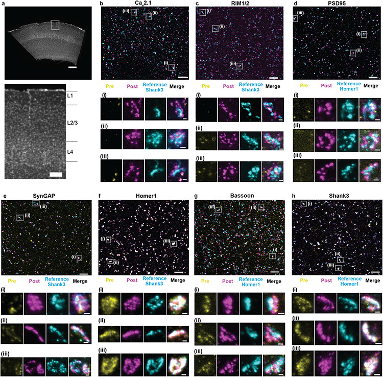
Fig. 3 ∣. Validation of ExR enhancement and effective resolution in synapses of mouse cortex.
(a) Low-magnification widefield image of a mouse brain slice with DAPI staining showing somatosensory cortex (top) and zoomed-in image (bottom) of boxed region containing L1, L2/3, and L4, which are imaged and analyzed after expansion further in panels b-h. (Scale bar, 300 μm (top) and 100 μm (bottom)). (b-h) Confocal images of (max intensity projections) of specimen after immunostaining with antibodies against Cav2.1 (Ca2+ channel P/Q-type) (b), RIM1/2 (c), PSD95 (d), SynGAP (e), Homer1 (f), Bassoon (g), and Shank3 (h), in somatosensory cortex L2/3. For pre-expansion staining, primary and secondary antibodies were stained before expansion, the stained secondary antibodies anchored to the gel, and finally fluorescent tertiary antibodies applied after expansion to enable visualization of pre-expansion staining. For post-expansion staining, the same primary and secondary antibodies were applied after ExR. Antibodies against Shank3 (b, c, e, f) or Homer1 (d, g, h) were applied post-expansion as a reference channel. Confocal images of cortex L2/3 (top) show merged images of pre- and post-expansion staining, and the reference channel. Zoomed-in images of three regions boxed in the top image (i-iii, bottom) show separate channels of pre-expansion staining (yellow), post-expansion staining (magenta), reference staining (cyan), and merged channel. (Scale bar, 1.5 μm (upper panel); 150 nm (bottom panel of i-iii).) Shown are images from one representative experiment from two independent replicates.

