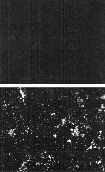
FIG. 2.
Detection of the 40-kDa protein (Emp) on the surface of S. aureus Newman by immunofluorescence microscopy. S. aureus cells were incubated with preimmune serum (top panel) or with antiserum directed against recombinant Emp preadsorbed with surface molecules from mutant mEmp50. Thereafter, bound immunoglobulins were detected by using FITC-labeled anti-IgG antibodies and examined by fluorescence microscopy. Magnification, ×200.

