
Figure 2.
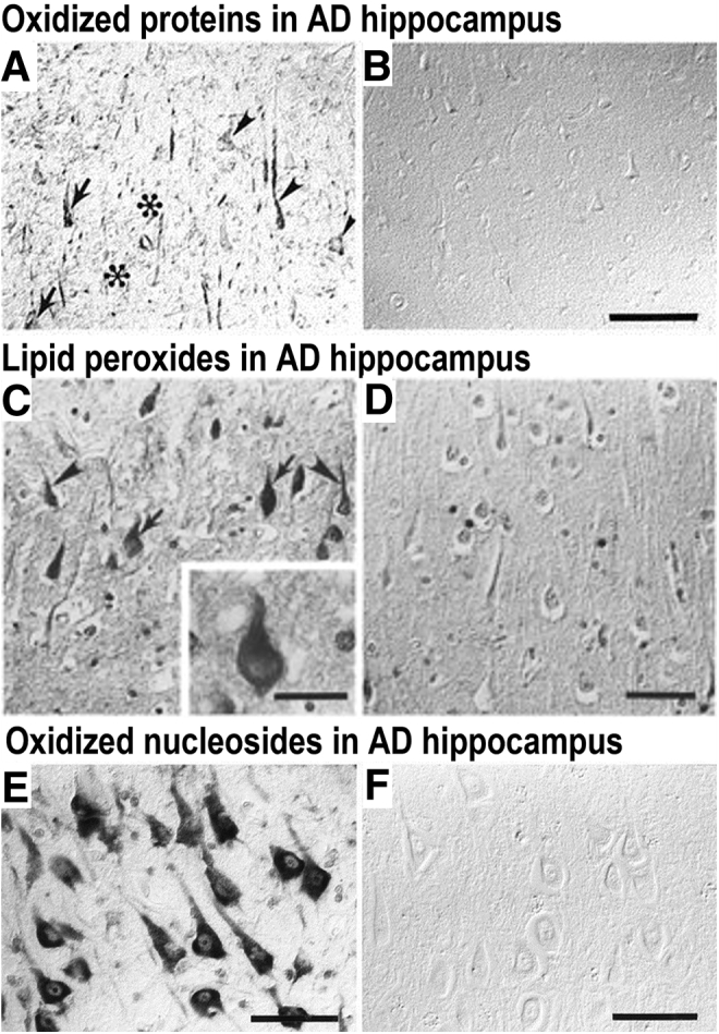
Oxidative stress in Alzheimer disease (AD). A: Immunolocalization of oxidized proteins (nitrotyrosine) in AD hippocampus [arrows indicate neurons with neurofibrillary tangles (NFTs), arrowheads indicate neurons without NFTs, and asterisks indicate location of senile plaques]. B: Control case (non-AD). C: Lipid peroxidation (4-hydroxy-2-nonenal–pyrrole) immunostaining in AD hippocampus (arrows indicate location of neurons with NFTs, and arrowheads indicate location of neurons without NFTs). Inset: A high-magnification view of a neuron containing an NFT and lipid peroxidation. D: Control (non-AD). E: Immunolocalization of oxidized nucleosides (anti–8-hydroxy-deoxyguanosine and anti–8-hydroxyguanine) in AD hippocampus. F: Control (non-AD). Reproduced with permission from J Neurosci, 1997, 17:2653–2657 (A and B)11; J Neurochem, 1997, 68:2092–2097 (C and D)12; and J Neurosci, 1999, 19:1959–1964 (E and F).13 Scale bars: 100 μm (B); 50 μm (C–F).