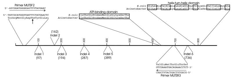
FIG. 3.
Conserved and divergent amino acid sequences in Acinetobacter MutS. Horizontal arrows indicate degenerate primers originally used to amplify a portion of mutS from the chromosome of strain ADP1. Boxes surround amino acid residues conserved in the Acinetobacter MutS and in the E. coli MutS for which the crystal structure has been determined (29). A vertical arrow indicates a conserved phenylalanine residue that has been shown to be required for mismatch binding in other MutS homologs. Numbers in parentheses indicate positions in the E. coli MutS sequence corresponding to the starts of indels that distinguish the primary sequences of the two MutS proteins.

