Fig. 2.

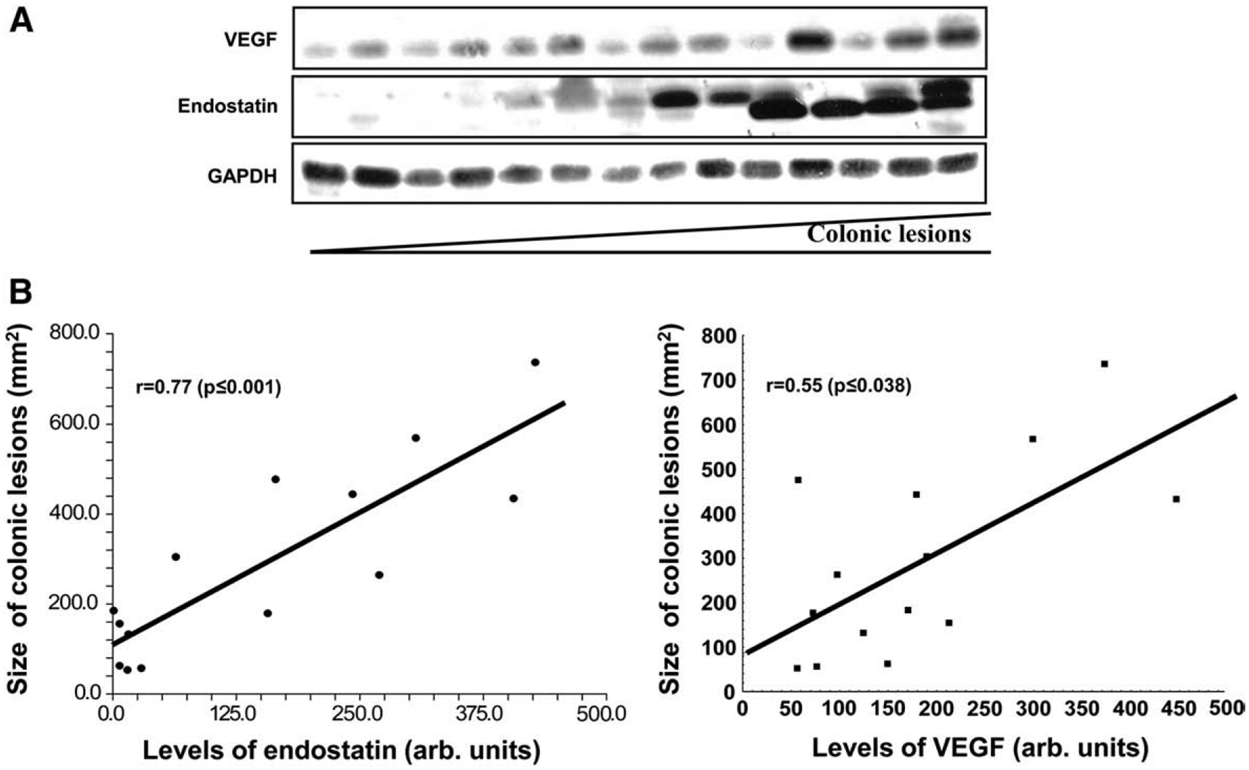
The correlation between colonic levels of endostatin, VEGF and size of colonic lesions during iodoacetamide-induced UC. (A) Protein levels in colonic tissue were determined by Western blot. Samples distributed from left to right according to size of colonic lesions from the smallest to greatest. GAPDH levels were used as loading controls. (B) Linear regression report and Spearman’s rank correlation coefficient. The density of Western blot bands was measured by the Eagle Eye II (Stratagene, Austin, TX) and presented in arbitrary units. Assays were repeated 3 times with highly reproducible results using protein from 3 rats/group.
