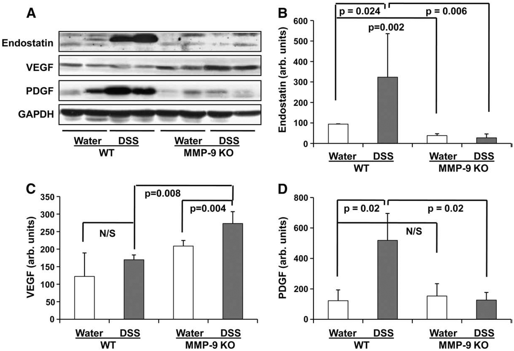
Fig. 5.

Levels of endostatin, angiostatin, VEGF and PDGF in colonic tissue of wild-type (WT) and MMP-9 KO mice with DSS-induced UC and water-ingested control. Protein levels in colonic tissue were determined by Western blot. A: Representative Western blot bands of endostatin, VEGF, and PDGF; B: Western blot density of endostatin; C: Western blot density of VEGF; D: Western blot density of PDGF. GAPDH levels were used as loading controls. Assays were repeated three times with highly reproducible results.
