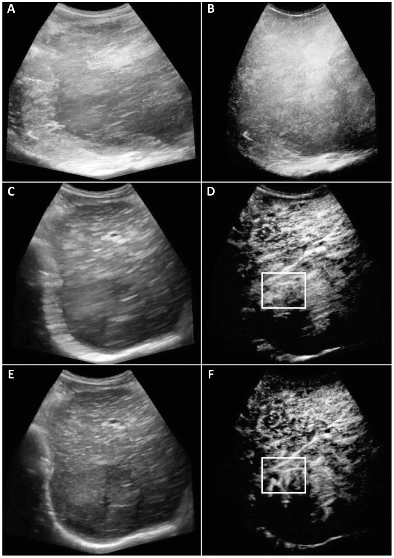
Fig. 2.

Original (A-B), out-of-plane frames eliminated (C-D), the resulting motion corrected (E-F) B-Mode (left) and CEUS (right) maximum intensity projection (MIP) images from 787, 255, and 255 frames respectively. Image size was 649×585 pixels. Highlighted changes in the white box did not contain any vessels for the original image while the corrected image shows the vessels, bifurcations, and tortuosity.
