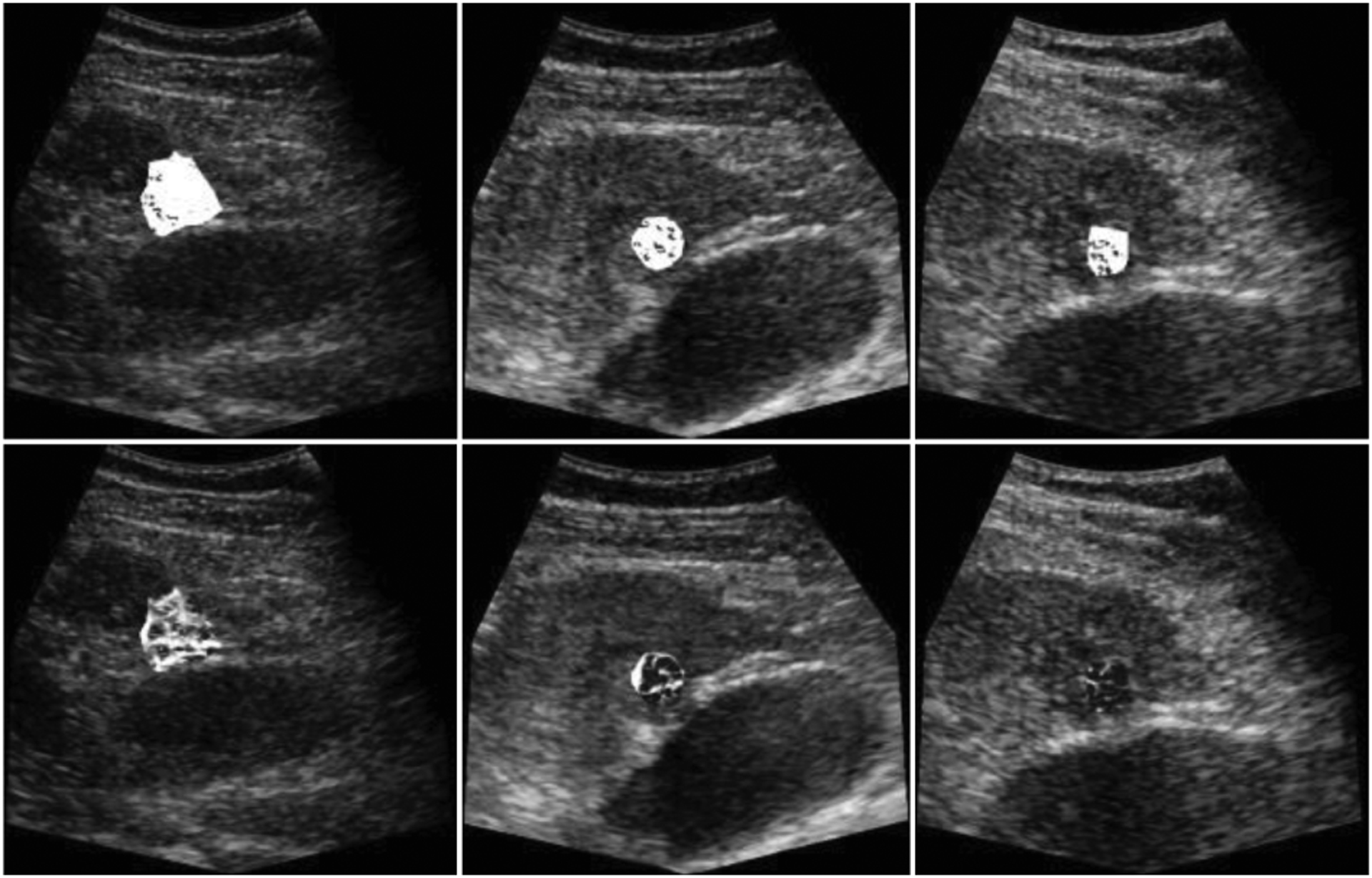
Fig. 3.

Vessel enhancement from region-of-interests (ROIs) were performed on the MIP of 255 frames and overlaid on a single B-Mode image for baseline (left), after two-wk (middle), and four-wk (right). The complete response from the clinical results were in line with the images (bottom). Dense vascularity on the baseline decreased after two- and four-wk. These temporal changes of the vascular morphology were not observable from the images (top) corrupted with motion.
