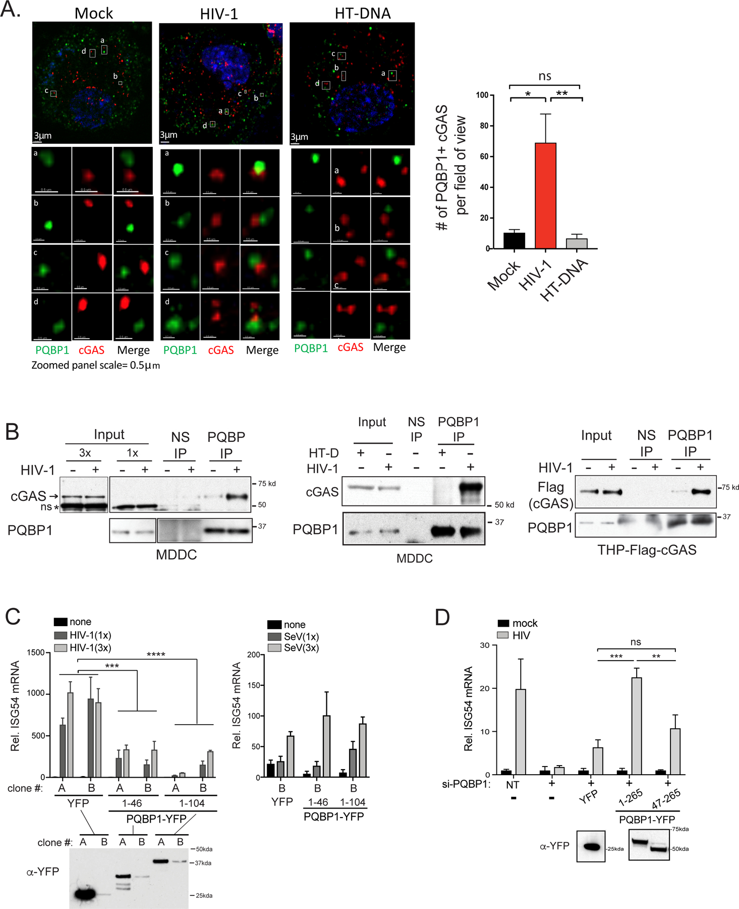
Figure 5. HIV-1 infection promotes the formation of a PQBP1/cGAS innate sensing complex.

HIV-1 infection enhances PQBP1 and cGAS interactions. (A) THP-1 cells treated with PMA were fixed 2.5 hours post HIV-1 infection (Gag-IN-mRuby) or HT DNA transfection, followed by immunostaining of PQBP1 and cGAS for confocal microscopy. Z-section images (left) and the quantification of coassociating foci (right) are shown. A threshold for colocalization is set at a distance of 0.4 µm. Averages and SEM are shown. One-way ANOVA, ** p<0.01, * p<0.05, ns denotes no significance. Scale bars: 3μm (top) and 0.5 μm (bottom). (B) HIV-1 infection (+) enhances coimmunoprecipitation of cGAS or Flag-cGAS with PQBP-IP in MDDCs or PMA-THP-1, respectively. Cells were infected with HIV-1 luciferase virus in the presence of VLP-Vpx and subjected to co-IPs at 3 hrs post-infection. HT-DNA was delivered to cells instead of viral infection, where indicated. Endogenous cGAS or Flag-cGAS co-precipitating with PQBP1 was assayed. 1x and 3 x denote the relative amounts of inputs loaded on a gel. Normal IgG (NS) or antibody against PQBP1 were used as indicated. ns denotes non-specific bands. Cropped images are enclosed by boxed frames. See Figure S5A for original uncropped images. (C) The capsid interaction domain of PQBP1 is a dominant inhibitor of cGAS-mediated innate sensing of HIV-1 infection. Two independent PMA-THP-1 cell clones (A and B clones) stably expressing either YFP, PQBP11–46-YFP or PQBP11–104-YFP were infected with HIV-1 luciferase virus in the presence of VLP-Vpx or Sendai virus as indicated. ISG54 mRNAs were measured 16 hours post infection by qRT-PCR. (D) The capsid interaction domain of PQBP1 is needed for the innate response against HIV-1 infection. THP-1 cells treated with PMA, either un-transduced or stably expressing the indicated PQBP1-YFP proteins, were treated with siRNA against endogenous PQBP1 (+), followed by HIV-1 infection and ISG54 mRNA detection as in (C). NT denotes non-targeting siRNA. ISG54 mRNA levels were expressed as fold-induction over their uninfected counterparts. Expression level of either YFP or PQBP-YFPs in the cells used in (C) and (D) were determined by anti-GFP/YFP western blots. Equal numbers of cells were analyzed. T-test (unpaired; two-tailed) *p<0.05, **p<0.01, ***p<0.001. All data, unless noted otherwise, are representative of at least two independent experiments. See also Figure S5.
