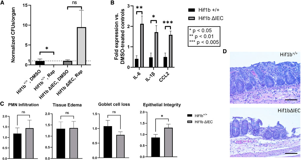
Figure 7. Treatment of mice with autophagy agonist RAP ameliorates STm-induced inflammation in a HIF-1β-dependent manner.
(A) Measurement of liver STm CFUs in mice treated either with DMSO or RAP. Data presented as CFUs per organ, normalized to DMSO-treated controls.
(B) Expression of pro-inflammatory cytokines in terminal ilea of mice infected with STm and treated with either DMSO or RAP. Data represented as fold expression of RAP-treated mice versus DMSO-treated controls.
(C) Histopathology of Salmonella-infected, RAP-treated Hif1b+/+ or Hif1bΔIEC mice was evaluated by H&E staining and microscopy. Data presented as total final score for indicated parameters.
(D) Representative histopathologic images of cecal tissue from Hif1bfl/fl (top) or Hif1bΔIEC (bottom) mice administered RAP in combination with STm. Note loss of epithelial integrity in Hif1bΔIEC. For all experiments, n = 9–10 mice per experimental group.
All statistics calculated by one-way ANOVA (A) or unpaired t test (B and C). *p < 0.05, **p < 0.01, ***p < 0.005 in all panels. Microscopy images at 400×; scale bar: 50 μm.

